1. Research on Microscopic Body Structure
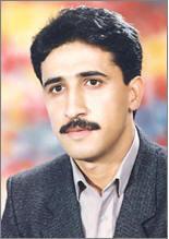
The head of Micro Anatomy Research Center of Mashhad University of Medical Sciences, Dr. Ebrahimzade said: research…
Created on 12 December 2016
The head of Micro Anatomy Research Center of Mashhad University of Medical Sciences, Dr. Ebrahimzade Bideskan said: research on the microscopic body structure is a research orientation in this center. According to WebDa, in a report on the 3-year practices of Micro Anatomy Research Center, Dr. Ebrahimzadeh spoke about the date of establishing this research center and said: the history of research here refers to not long ago as the activities here have been started in 2014.
“Micro Anatomy Research Center has a well-equipped laboratory in which major research and scientific investigations are done with student participation”, he added.
The head of Micro Anatomy Research Center pointed out: this center is built in Medical School funded through the collaboration between this center and private sector with the participation of charitable philanthropists. The required equipment here have been purchased by the university within a period of four years and handed in to the researchers in this center.
Dr. Ebrahimzadeh averred that this center was established with the private sector and altruists’ sponsorship with an expense of 10,000 dollars. The university purchased the equipment along a period of four years.
Referring to active members in research and experiment in this center, he highlighted: major activities here are undertaken by faculty members of educational departments of anatomical sciences, cellular biology, biochemistry and other educational groups.
The head of Micro Anatomy Research Center asserted: a significant purpose of establishing this center has been the presence and participation of capable and active members in addition to creating links among basic and clinical sciences in the scope of histology and pathology.
“As micro anatomy work is done in the scope of microscopic anatomy science, we attempted to create an interdisciplinary collaboration and cooperation among the specialists who share concerns in this regard”, he said.
Dr. Ebrahimzadeh stated: microscopic anatomy used to be presented in the course of anatomical sciences and its pathology by our colleagues in pathology department. We attempted to finally strengthen the relation between these two scopes through the establishment of this research center.
The head of Micro Anatomy research Center stressed: this center is now in early years of its activities, as only one and a half years have passed from its official establishment. Though in this short while, we have been able to present an equivalent of three articles for each active educational faculty member here.
He considered the collaboration of basic and clinical science specialists as a leading cause of scientific development and production in research centers, and highlighted: in most cases we observe that clinical specialists are interested in participating in the scopes of basic sciences. There is such a possibility to have successful projects based upon the ideas that these clinical specialists may suggest.
Speaking of the inauguration ceremony of Neurosciences Department in Medical School last month, he stressed: at the moment, various groups of researchers participate and pursue their experimental research activities in this center. Recently, with the inauguration of Neurosciences Department in this school, we are seeking to establish favorable collaboration and cooperation in interdisciplinary sciences.
The head of Micro Anatomy Research Center asserted: as this center is in initial condition of its activities, and pursues its major projects focused on student theses, it needs some time till the success targeted by the university seems visible.
Highlighting the importance of presenting advanced ideas in research centers, he said: in case good ideas are presented among research centers, high levels of successful achievements can be obtained through building the connection among these centers, which is a concern to be followed in the pursuit of activities in this research center.
The head of Micro Anatomy Research Center asserted: here, microscopic anatomy and the cellular and texture structure and construction of body tissues are investigated, and there are efforts to concentrate on those cellular structures that can be seen through microscope.
In the end, Dr. Ebrahimzadeh stated: capable human resources shape the foundation of each association. Accordingly, with the support of authorities in the university, we hope to be one of the prominent research centers of Mashhad University of Medical Sciences in the years to come.
2. 3D Printing Modern Technology
... “This method is used in anatomical trainings, clinical therapies and surgery”. The head of Radiology Department of Mashhad University of Medical Sciences noting that various departments of the university ...
Created on 31 January 2016
According to WEBDA, this technology is being studied by Dr. Sirous Nekouyi, head of Radiology Department of Mashhad University of Medical Sciences, in the form of research projects, and effective measures have been taken so far in this area. Stating that currently 3D printing has a wide range of applications in industry and in making industrial models and tools, Dr. Sirous Nekouyi said: “This technology is being used as a new method in the field of education and treatment in medical sciences in developed countries. This faculty member of Mashhad University of Medical Sciences noted: “In this method, data is extracted using the sophisticated technology of the results of CT scan and MRI, and the desired part is made. Dr. Nekouyi added: “After feeding data into the machine, the desired sample is heated using plastic materials, and leaves the machine nozzle in the form of strings in layers to a thickness of a tenth of a millimeter. Stating that the duration of printing each part was one to two hours, He explained: “This method is used in anatomical trainings, clinical therapies and surgery”.
The head of Radiology Department of Mashhad University of Medical Sciences noting that various departments of the university such as the Neurology Department have so far used this method in surgeries stated: “This method is used in surgery in such a way that the desired organ is initially made in a 3D form, and then the surgery is held after examining certain injuries”. Dr. Nekouyi Stating that this method is best applicable in the field of anatomy said: “Currently, students study different body organs through books or computers while different body organs, tissues, arteries, neurons and vessels can be tangibly taught to students using this method”.

.jpg)
